Knee Surgery / Arthroscopic Multiligament Reconstruction
Arthroscopic Multiliagment reconstruction is commonly known as keyhole surgery which allows the surgeon to view the inside of your joint, providing a more accurate way to diagnose and treat certain … conditions.
What is Arthroscopic Multiliagment Reconstruction
A small camera is inserted into the knee and the images are relayed to a television screen.
Specialist instruments are introduced into the joint through small incisions (less than 1cm). Using these instruments a surgeon can perform a number of procedures. These include washing out fluid or tissue debris, removing or reattaching loose fragments, repairing or removing torn cartilage.
Reason for An Arthroscopic Multiliagment Reconstruction
- An Arthroscopic Multiliagment reconstruction may be done to investigate symptoms (diagnostic) such as pain, swelling, or instability of a joint.It may show damage to cartilage or ligaments within a joint, fragments of bone or cartilage which have broken off (‘loose bodies’), or signs of arthritis.
- The menisci are 2 semi- circular structures of soft fibro-cartilage which act as shock absorbers within the joint. They are often injured by twisting activities. If you have a tear in the meniscus the torn section is ‘trimmed’ back to healthy stable meniscus. Often,it is possible to repair the torn cartilage, although this is most common in the young adult or child
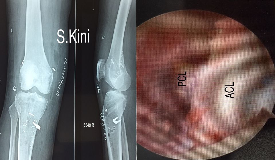
- The Cruciate ligaments are 2 strong ligaments, the anterior and the posterior, which provide stability of the knee on twisting and pivoting activities. They are often injured in contact sports and skiing. Arthroscopy allows a clear view and physical inspection of the Cruciate ligaments and gives us the opportunity to reconstruct them if necessary.
- Small fragments of bone or articular cartilage can become loose within the knee joint often through trauma or degenerative changes (osteoarthritis). These can be removed and ‘washed out’ of the joint.
- An arthroscopy can assess the extent of damage that has occurred when a knee is injured eg. The smooth articular cartilage lining of the bone which allows smooth movement may be damaged when the knee is injured, this can result in a ‘divot’ of cartilage becoming loose and causing pain and locking of the joint. The cartilage surface may be treated in a number of ways, including microfracture….
- If the joint lining is particularly inflamed, a small area of this lining can be taken (a biopsy) and sent for further investigations as to the cause.
- An arthroscopy can be used to remove /release scar tissue. For example, in very specific cases, a procedure called a lateral release can be performed. This is the surgical division of the soft tissues on the outer aspect of the patella. These structures can be extremely tight causing the patella to track in the wrong position. This tightness over a long time can place excess pressure on the under surface of the patella resulting in pain.
- The kneecap (patella) can be a source of pain in the knee. The arthroscope allows inspection of the patella under surface. If there is any loose articular cartilage this can be shaved.